Brain Anatomy in Psychiatric Disorders
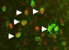
The image above is an example of mapping cellular responses to stress. To produce mild stress, a rat was given an injection of saline (saline is water with the normal concentration of blood salt and does not disturb any function in the body). The image above was taken from the lateral septum, a brain region that is known to respond to stress. One of our objectives is to understand cellular responses to stress. In the image above, a subset of cells express both FOS (red), and acetylcholinesterase (green) proteins (see white arrow-heads). We have applied this type of staining to all brain regions, as part of building a map of coordinated cellular responses to stressful experiences.
Brain systems that organize the response to a threatening situation appear to have two facets – one is emotional, e.g. the experience of fear, anxiety, or even panic and the other is motor, e.g. “freezing”, exemplified by complete immobility, or escape, as manifested in running, or as appropriate under some conditions, fighting.
Since the brain systems controlling reactions to stressful situations, employ neurotransmitters, imbalance between neurotransmitter systems may underlie abnormal responses to psychological stress. In collaboration with Prof. Hermona Soreq from the Hebrew University in Jerusalem, we focus the cholinergic system and its interaction with other systems. One facet of cholinergic mechanisms is the alternative splicing of acetylcholinesterase. A new and exciting field is emerging based on the demonstration that one splice variant, the acetylcholinesterase readthrough (AChE-R), is increased in brain of mice after exposure to acute stress. To study the impact of AChE-R on behavior, a line of transgenic mice has been prepared that has increased expression of human AChE-R.
We have mapped the distribution of AChE-R in these mice and found it in brain systems that enhance fearful behavior and in systems that keep fearful behavior under check (Sternfeld et al 2000, Birikh et al 2001). Subsequent work has shown that AChE-R synthesis in the hippocampus participates in fear-motivated learning (Nijholt et al. 2003). This work may lead to insights into Posttraumatic stress disorder (PTSD).
Interactions between emotional and motor systems in psychiatric disorders
Abnormal motor acts have been observed in a wide variety of psychiatric disorders. Although the underlying mechanisms have been attributed to basal ganglia and/or cerebellar systems and their interactions with cortical brain regions, it is not clear what neurotransmitter pathways may be involved and how they should be manipulated in a way that would normalize motor behavior without unwanted side effects.
In the case of schizophrenia, motor hyperactivity and impaired cognition have been modeled by phencyclidine injection to mice and/or monkeys. The amino acid glycine acts as an inhibitory neurotransmitter in brain systems controlling motor behavior and also modulates transmission by an excitatory amino acid, glutamate. In collaboration with Dr. Uriel Heresco-Levy from Herzog Hospital and Dr. Dan Javitt from the Nathan Kline Institute in Ellensburg, New York, we have studied the impact of glycine and glycine-related compounds on brain and on motor behavior (Shoham et al 1999, 2001, and 2004 in press). These studies may lead to new approaches for treatment of motor abnormalities in both psychiatric and neurological disorders.
In several psychiatric disorders there are stereotypic behaviors. Namely repetitive, apparently purposeless motor behaviors. In collaboration with Prof. Hermona Soreq of the Hebrew University in Jerusalem, we are studying the mechanisms underlying stereotypic behavior in a transgenic mouse model (Shoham et al 2002).


